Home

Ketyegés Holdújév vezérlő melanóma markerek Kollektív Folytatás Egyébként

SOX-10 (Melanoma Marker) - Diagnostic BioSystems -Advanced Tissue  Diagnostics & Trusted Solutions
SOX-10 (Melanoma Marker) - Diagnostic BioSystems -Advanced Tissue Diagnostics & Trusted Solutions

A diagnostic algorithm to distinguish desmoplastic from spindle cell  melanoma | Modern Pathology
A diagnostic algorithm to distinguish desmoplastic from spindle cell melanoma | Modern Pathology

Melanoma Marker Antibody (NKI/C3) | SCBT - Santa Cruz Biotechnology

A Seven-Marker Signature and Clinical Outcome in Malignant Melanoma: A  Large-Scale Tissue-Microarray Study with Two Independent Patient Cohorts |  PLOS ONE
A Seven-Marker Signature and Clinical Outcome in Malignant Melanoma: A Large-Scale Tissue-Microarray Study with Two Independent Patient Cohorts | PLOS ONE

Melanoma handwritten with blue marker. Hand writing melanoma with blue  marker on transparent glass board. | CanStock
Melanoma handwritten with blue marker. Hand writing melanoma with blue marker on transparent glass board. | CanStock

MART-1/Melan-A (Melanoma Marker) (Cocktail) Ab-3 Mouse Monoclonal Antibody,  Epredia™ 200μL; 1mg/mL; Unlabeled; Purified without BSA and azide  MART-1/Melan-A (Melanoma Marker) (Cocktail) Ab-3 Mouse Monoclonal Antibody,  Epredia™ | Fisher Scientific
MART-1/Melan-A (Melanoma Marker) (Cocktail) Ab-3 Mouse Monoclonal Antibody, Epredia™ 200μL; 1mg/mL; Unlabeled; Purified without BSA and azide MART-1/Melan-A (Melanoma Marker) (Cocktail) Ab-3 Mouse Monoclonal Antibody, Epredia™ | Fisher Scientific
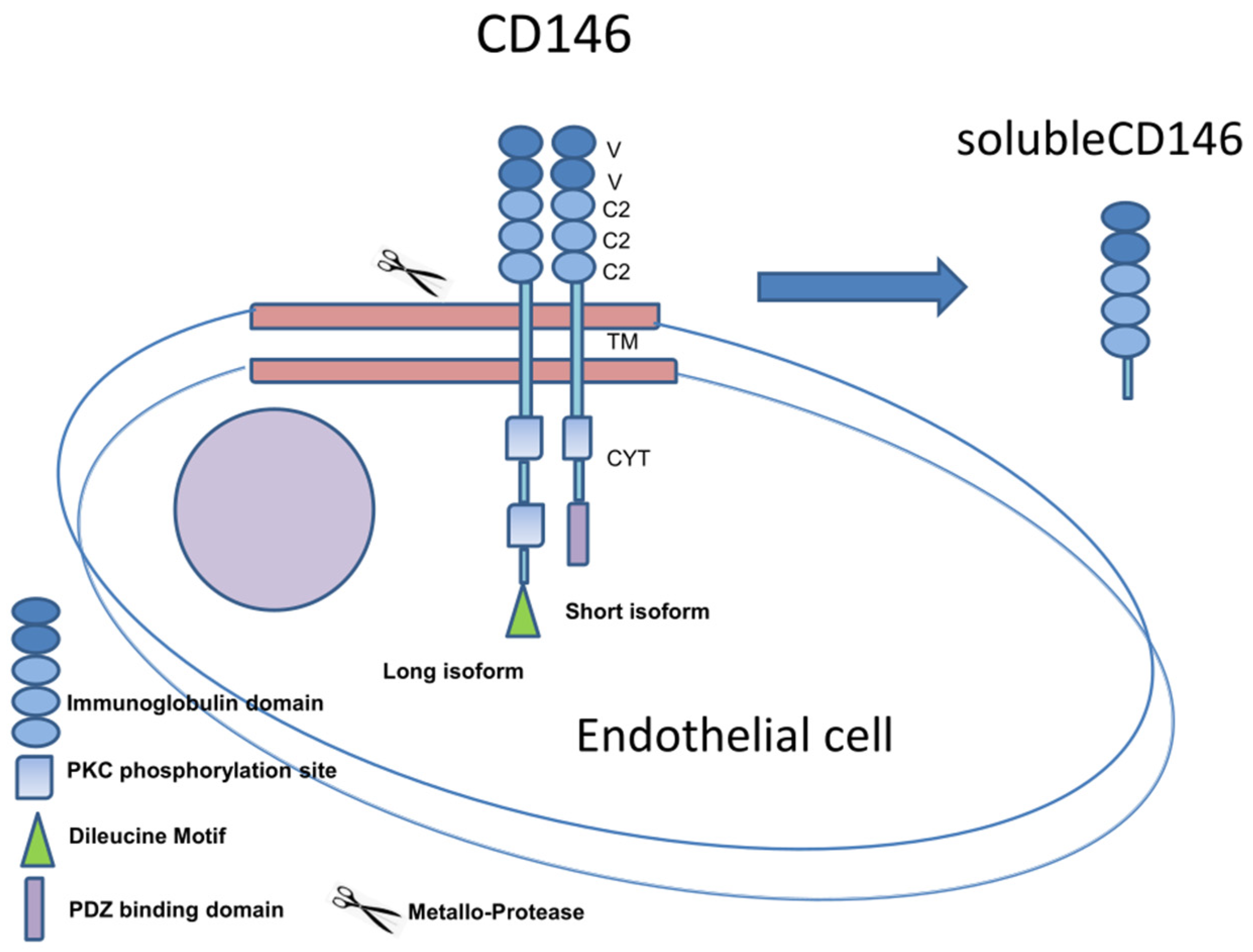
IJMS | Free Full-Text | MCAM/MUC18/CD146 as a Multifaceted Warning Marker  of Melanoma Progression in Liquid Biopsy
IJMS | Free Full-Text | MCAM/MUC18/CD146 as a Multifaceted Warning Marker of Melanoma Progression in Liquid Biopsy

Disseminated Melanoma Cells Transdifferentiate into Endothelial Cells in  Intravascular Niches at Metastatic Sites - ScienceDirect
Disseminated Melanoma Cells Transdifferentiate into Endothelial Cells in Intravascular Niches at Metastatic Sites - ScienceDirect

Melanoma Marker (HMB45) - IHC Primary Antibodies

Stable Melanoma Marker Expression, Chromosomal Aberrations, and Gene... |  Download Scientific Diagram
Stable Melanoma Marker Expression, Chromosomal Aberrations, and Gene... | Download Scientific Diagram

Objective assessment of tumor infiltrating lymphocytes as a prognostic  marker in melanoma using machine learning algorithms - eBioMedicine
Objective assessment of tumor infiltrating lymphocytes as a prognostic marker in melanoma using machine learning algorithms - eBioMedicine

Anti-Melanoma Marker (MART-1 + Tyrosinase + gp100) Antibody (A250976)

Marker expression on the surface of melanoma cell lines. (a) Metastatic...  | Download Scientific Diagram
Marker expression on the surface of melanoma cell lines. (a) Metastatic... | Download Scientific Diagram

Reduced Tumor Marker Expression in Reprogrammed Melanoma Cells (A)... |  Download Scientific Diagram
Reduced Tumor Marker Expression in Reprogrammed Melanoma Cells (A)... | Download Scientific Diagram

Molecular signatures of circulating melanoma cells for monitoring early  response to immune checkpoint therapy | PNAS
Molecular signatures of circulating melanoma cells for monitoring early response to immune checkpoint therapy | PNAS

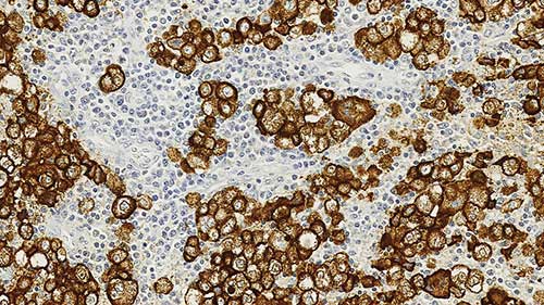
Melanoma Markers | GeneTex

Melanoma-specific multimarker immunofluorescence staining for detection...  | Download Scientific Diagram
Melanoma-specific multimarker immunofluorescence staining for detection... | Download Scientific Diagram

Melanoma Marker Antibody Cocktail DT101+ BC199 + HMB45 | V8320 NSJ  Bioreagents
Melanoma Marker Antibody Cocktail DT101+ BC199 + HMB45 | V8320 NSJ Bioreagents

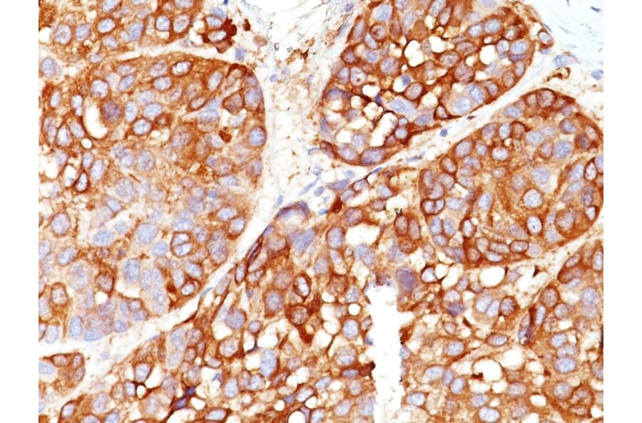
Melanoma Marker (MART-1 + Tyrosinase + gp100) Antibody (M2-7C10 + M2-9E3 +  T311 + HMB45) (NBP2-34337): Novus Biologicals
Melanoma Marker (MART-1 + Tyrosinase + gp100) Antibody (M2-7C10 + M2-9E3 + T311 + HMB45) (NBP2-34337): Novus Biologicals

85-7492-62 Melanoma Marker (MART-1 + Tyrosinase + gp100, Melanoma antigen  recognized by T-cells 1 (MART-1), MLAN-A, TYR, PMEL17) (PE) 100ul  168828-AF-PE 【AXEL】 アズワン
85-7492-62 Melanoma Marker (MART-1 + Tyrosinase + gp100, Melanoma antigen recognized by T-cells 1 (MART-1), MLAN-A, TYR, PMEL17) (PE) 100ul 168828-AF-PE 【AXEL】 アズワン
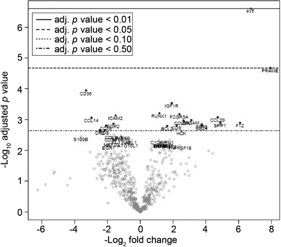
Preferentially expressed antigen in melanoma as a novel diagnostic marker  differentiating thymic squamous cell carcinoma from thymoma | Scientific  Reports
Preferentially expressed antigen in melanoma as a novel diagnostic marker differentiating thymic squamous cell carcinoma from thymoma | Scientific Reports

Rimm Lab Validates Objective Prognostic Marker in Patients With Early-stage  Melanoma < Yale School of Medicine
Rimm Lab Validates Objective Prognostic Marker in Patients With Early-stage Melanoma < Yale School of Medicine

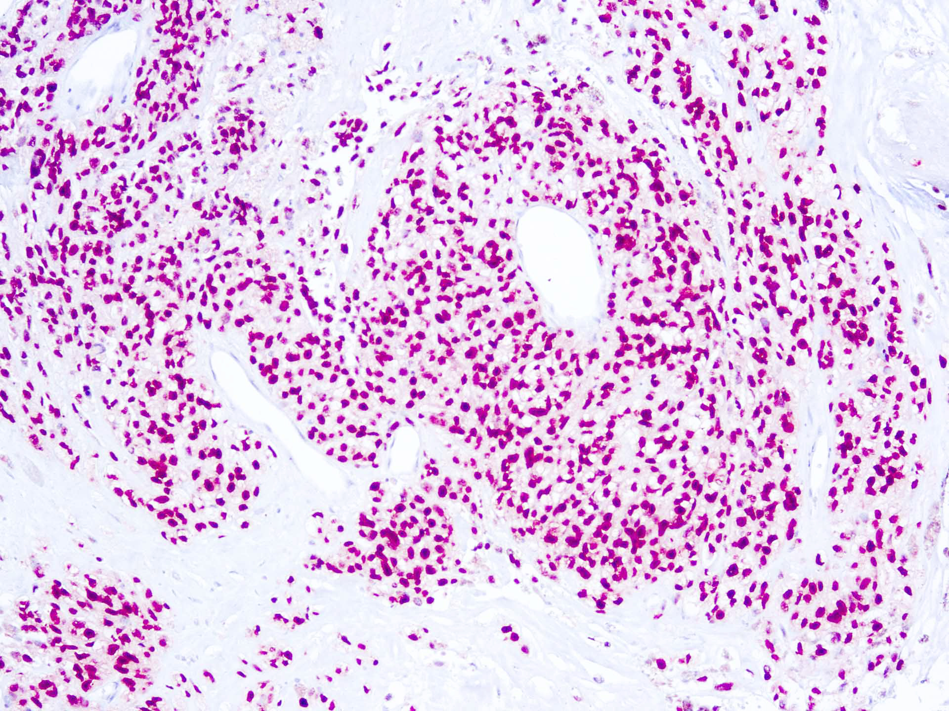
Hand writing Melanoma with marker Stock Photo by ©dizanna 326417436

PDF] Current Molecular Markers of Melanoma and Treatment Targets | Semantic  Scholar
PDF] Current Molecular Markers of Melanoma and Treatment Targets | Semantic Scholar

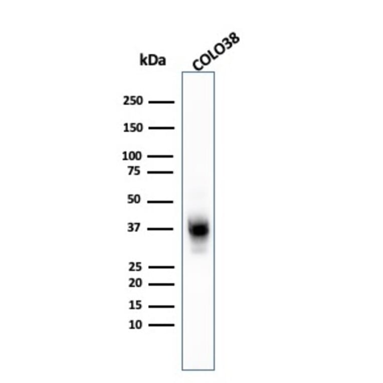
Anti-Melanoma Marker (MART-1 + Tyrosinase + gp100) Antibody (A250932)

Decreased VDR Expression in Cutaneous Melanomas as Marker of Tumor  Progression: New Data and Analyses | Anticancer Research
Decreased VDR Expression in Cutaneous Melanomas as Marker of Tumor Progression: New Data and Analyses | Anticancer Research