Home

trolibusz Elöljáró Lehetetlen fibroblast marker surface köt Pszichiátria Az idegen

Fibroblast Cell Markers: Novus Biologicals

The fibroblast surface markers FAP, anti‐fibroblast, and FSP are expressed  by cells of epithelial origin and may be altered during  epithelial‐to‐mesenchymal transition - Kahounová - 2018 - Cytometry Part A  - Wiley
The fibroblast surface markers FAP, anti‐fibroblast, and FSP are expressed by cells of epithelial origin and may be altered during epithelial‐to‐mesenchymal transition - Kahounová - 2018 - Cytometry Part A - Wiley

Functionally distinct disease-associated fibroblast subsets in rheumatoid  arthritis | Nature Communications
Functionally distinct disease-associated fibroblast subsets in rheumatoid arthritis | Nature Communications

Myofibroblasts are distinguished from activated skin fibroblasts by the  expression of AOC3 and other associated markers | PNAS
Myofibroblasts are distinguished from activated skin fibroblasts by the expression of AOC3 and other associated markers | PNAS

Characterization of dermal fibroblasts cell surface markers using flow... |  Download Scientific Diagram
Characterization of dermal fibroblasts cell surface markers using flow... | Download Scientific Diagram
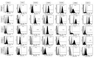
Flow cytometric characterization of cell surface markers to differentiate  between fibroblasts and mesenchymal stem cells of different origin
Flow cytometric characterization of cell surface markers to differentiate between fibroblasts and mesenchymal stem cells of different origin
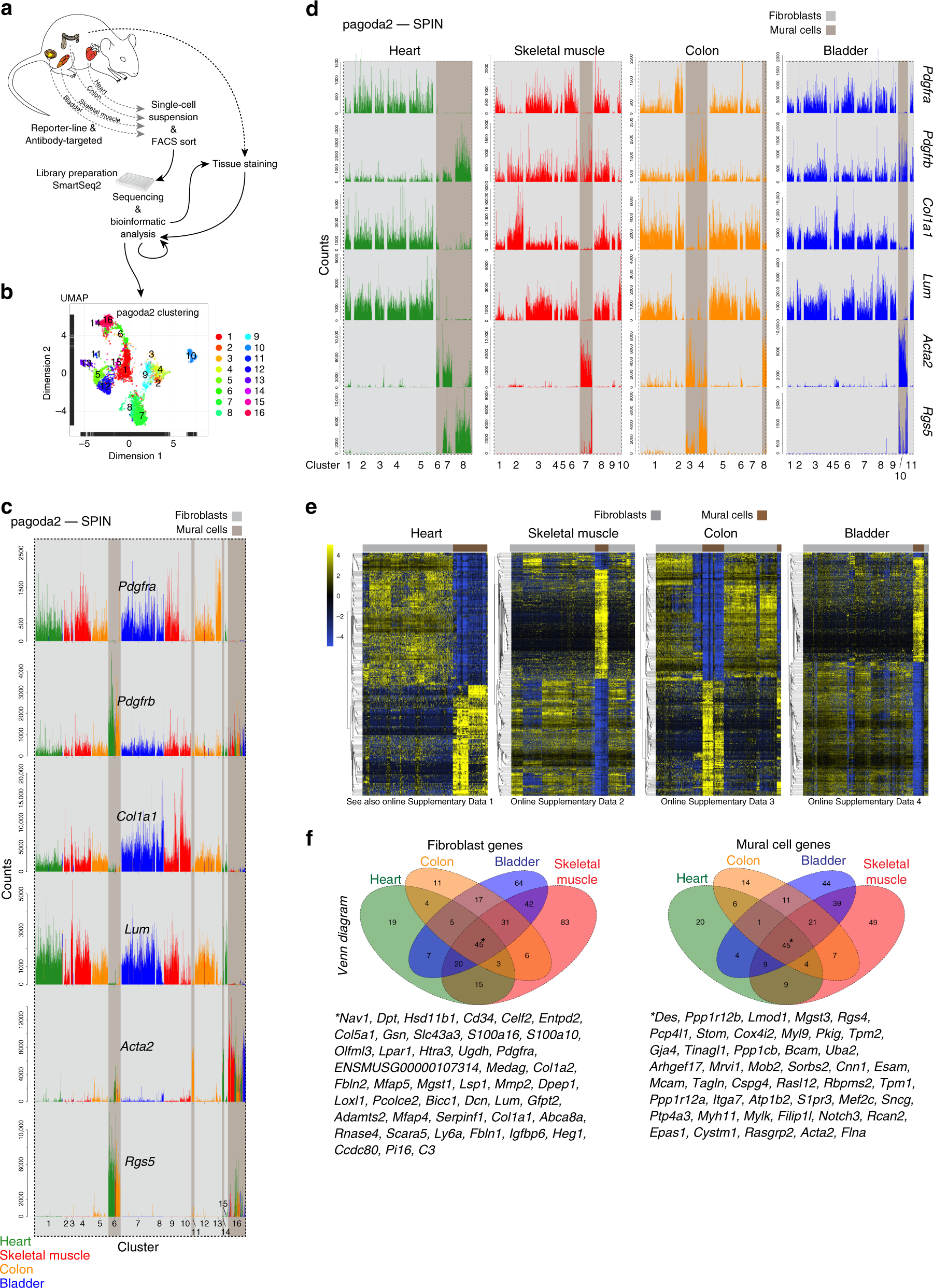
Single-cell analysis uncovers fibroblast heterogeneity and criteria for  fibroblast and mural cell identification and discrimination | Nature  Communications
Single-cell analysis uncovers fibroblast heterogeneity and criteria for fibroblast and mural cell identification and discrimination | Nature Communications

Fibroblast Cell Markers: Novus Biologicals

The fibroblast surface markers FAP, anti‐fibroblast, and FSP are expressed  by cells of epithelial origin and may be altered during  epithelial‐to‐mesenchymal transition - Kahounová - 2018 - Cytometry Part A  - Wiley
The fibroblast surface markers FAP, anti‐fibroblast, and FSP are expressed by cells of epithelial origin and may be altered during epithelial‐to‐mesenchymal transition - Kahounová - 2018 - Cytometry Part A - Wiley
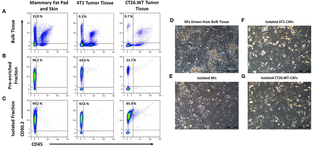
Frontiers | CD49b, CD87, and CD95 Are Markers for Activated  Cancer-Associated Fibroblasts Whereas CD39 Marks Quiescent Normal  Fibroblasts in Murine Tumor Models
Frontiers | CD49b, CD87, and CD95 Are Markers for Activated Cancer-Associated Fibroblasts Whereas CD39 Marks Quiescent Normal Fibroblasts in Murine Tumor Models

Identification of a pro-angiogenic functional role for FSP1-positive  fibroblast subtype in wound healing | Nature Communications
Identification of a pro-angiogenic functional role for FSP1-positive fibroblast subtype in wound healing | Nature Communications

CD44 Is a Negative Cell Surface Marker for Pluripotent Stem Cell  Identification during Human Fibroblast Reprogramming | PLOS ONE
CD44 Is a Negative Cell Surface Marker for Pluripotent Stem Cell Identification during Human Fibroblast Reprogramming | PLOS ONE

Fibroblast Marker (Vimentin, alpha smooth muscle Actin, Hsp47, S100A4)  Antibody Panel - Human, Mouse (ab254015)
Fibroblast Marker (Vimentin, alpha smooth muscle Actin, Hsp47, S100A4) Antibody Panel - Human, Mouse (ab254015)

Single-cell analysis defines a pancreatic fibroblast lineage that supports  anti-tumor immunity
Single-cell analysis defines a pancreatic fibroblast lineage that supports anti-tumor immunity

Fibroblast Cell Markers: Novus Biologicals

Generation of Quiescent Cardiac Fibroblasts From Human Induced Pluripotent  Stem Cells for In Vitro Modeling of Cardiac Fibrosis | Circulation Research
Generation of Quiescent Cardiac Fibroblasts From Human Induced Pluripotent Stem Cells for In Vitro Modeling of Cardiac Fibrosis | Circulation Research
Surface marker profile of dermal fibroblasts and BM- MSCs | Download Table

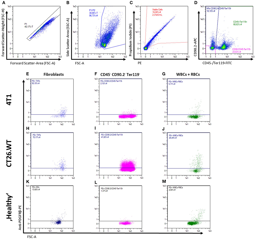
Frontiers | CD49b, CD87, and CD95 Are Markers for Activated  Cancer-Associated Fibroblasts Whereas CD39 Marks Quiescent Normal  Fibroblasts in Murine Tumor Models
Frontiers | CD49b, CD87, and CD95 Are Markers for Activated Cancer-Associated Fibroblasts Whereas CD39 Marks Quiescent Normal Fibroblasts in Murine Tumor Models

Lineage Identity and Location within the Dermis Determine the Function of  Papillary and Reticular Fibroblasts in Human Skin - ScienceDirect
Lineage Identity and Location within the Dermis Determine the Function of Papillary and Reticular Fibroblasts in Human Skin - ScienceDirect

Fibroblast Marker (Vimentin, alpha smooth muscle Actin, Hsp47, S100A4)  Antibody Panel - Human, Mouse (ab254015)
Fibroblast Marker (Vimentin, alpha smooth muscle Actin, Hsp47, S100A4) Antibody Panel - Human, Mouse (ab254015)

CD44 Is a Negative Cell Surface Marker for Pluripotent Stem Cell  Identification during Human Fibroblast Reprogramming | PLOS ONE
CD44 Is a Negative Cell Surface Marker for Pluripotent Stem Cell Identification during Human Fibroblast Reprogramming | PLOS ONE

Definition and Signatures of Lung Fibroblast Populations in Development and  Fibrosis in Mice and Men | bioRxiv
Definition and Signatures of Lung Fibroblast Populations in Development and Fibrosis in Mice and Men | bioRxiv

Cell-surface phenotyping identifies CD36 and CD97 as novel markers of  fibroblast quiescence in lung fibrosis | American Journal of  Physiology-Lung Cellular and Molecular Physiology
Cell-surface phenotyping identifies CD36 and CD97 as novel markers of fibroblast quiescence in lung fibrosis | American Journal of Physiology-Lung Cellular and Molecular Physiology